
The UMCG, located on the Healthy Ageing Campus, is the first hospital in the Netherlands to start a clinical study in which surgeons can immediately check during an operation whether a tumor has been completely removed. This is done using a mobile PET-CT scanner that scans the removed tissue directly in the operating room. In the future, this approach may help prevent a second surgery or additional treatments such as radiotherapy or chemotherapy.
Patients participating in the study receive a radioactive tracer before surgery. This tracer contains radioactive “sugar.” Because tumor cells have a higher sugar metabolism, they absorb more of the tracer than healthy cells. As a result, tumor cells become clearly visible on the PET-CT scan.
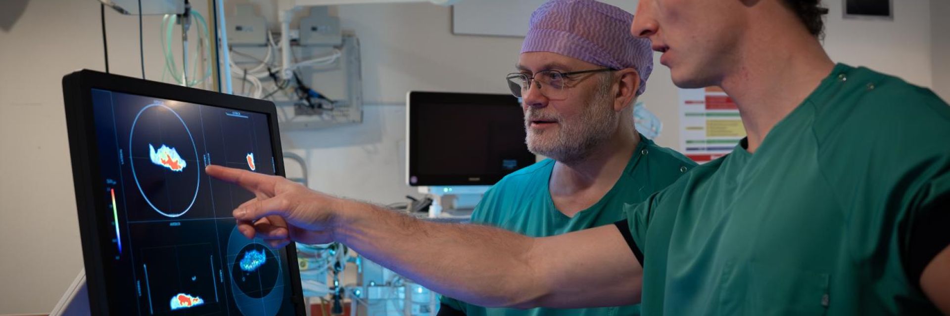
Immediately after the surgeon removes the tumor, the extracted tissue is scanned on the spot. The surgeon and the nuclear medicine specialist then assess together whether the surgical margins contain only healthy tissue. If tumor tissue is still visible, the surgeon can remove additional tissue during the same procedure.
This approach could save significant time in the future and may prevent a second operation or additional treatments such as radiotherapy or chemotherapy. Currently, the removed tumor tissue is examined by a pathologist to determine whether the margins still contain tumor cells. This process takes five to seven days. If the margins are not clear, the patient may need another operation or additional treatment. “A patient’s prognosis is always best when the tumor is completely removed in one go,” says researcher and radiology resident Jasper Vonk.
"This marks an important step toward making surgeries safer and more effective, and improving care for cancer patients."
Demonstrating Safety First
The first goal of the study is to demonstrate that it is safe to work with radioactive substances in the operating room. Staff therefore use radiation detectors, double gloves, and protocols for the safe disposal of radioactive patient materials and instruments used during surgery. Initial measurements indicate that this can be done safely, and early data from other countries show a similar picture.
The study, which includes thirty patients, will compare the PET images with the final assessment made by the pathologist. The PET-CT images are not yet being used to make decisions during surgery. That will only be possible in a next phase, once it is clear that the method is sufficiently reliable.
The Growing Role of Imaging in Surgery
The researchers expect that this technique will be especially beneficial for tumors located deep within the body or in cases where a complex operation is required. In the future, the technique may potentially be applied across all oncological surgery, as many tumors can be visualized using this PET tracer. In addition, there are other, more specific PET tracers available for different types of tumors.
The study demonstrates how imaging is becoming increasingly important during surgery. In the future, a technique like this could become a standard component of so‑called hybrid operating rooms, where advanced imaging is used directly during surgery to support surgeons.
Collaboration in the Operating Room
Close collaboration between nuclear medicine, the radiation protection unit, and surgery is essential for this approach. “This study brings colleagues who often still work at a distance from one another closer together. The PET scanner is literally coming into the operating room,” says Vonk.
“The concept of the image‑guided operating room can truly become reality with this,” says maxillofacial oncologic surgeon Max Witjes, who is involved in the study. “It represents an important step toward making surgeries safer and more effective, and improving care for cancer patients. And in a highly innovative way: the use of imaging techniques in the operating room is being tested in only a few places worldwide.”
Mandema Fellowship
Jasper Vonk is carrying out this project with a Mandema Fellowship, a personal grant from the UMCG that enables young, recently graduated physician‑researchers (residents in training) to combine their medical specialist training with establishing their own research line.
Source: UMCG, Marjolein te Winkel